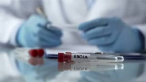
The Discovery of New Ebola Drug Targets Using ML-Powered Imaging
Introduction
Ebola virus disease (EVD) remains one of the most lethal viral infections known to humanity, with case fatality rates ranging from 25% to 90% depending on outbreak conditions. Despite progress in vaccine development and supportive care, effective antiviral drugs targeting Ebola remain limited. The urgent need for therapeutic interventions has driven scientists to explore innovative ways to identify potential drug targets within the virus’s life cycle.
One of the most promising approaches in recent years has been the integration of machine learning (ML) with advanced imaging technologies. By analyzing massive datasets of viral proteins, host–virus interactions, and cellular imaging, ML-powered imaging techniques are enabling researchers to uncover hidden patterns and discover novel therapeutic targets.
This article provides a deep dive into how machine learning combined with imaging is accelerating the discovery of new Ebola drug targets, the breakthroughs achieved so far, and the implications for future outbreaks and pandemic preparedness.
Ebola Virus: A Brief Overview
To understand the significance of these discoveries, it’s important to revisit the basics of Ebola virus biology.
-
Family and Structure: Ebola virus belongs to the Filoviridae family and is an enveloped, filamentous virus with a negative-sense single-stranded RNA genome.
-
Key Proteins: The viral genome encodes several proteins including glycoprotein (GP), nucleoprotein (NP), RNA polymerase (L), matrix proteins (VP24, VP35, VP40), and others.
-
Infection Mechanism: Ebola enters host cells primarily through macropinocytosis, mediated by the glycoprotein, and manipulates host immune responses to enhance replication.
-
Drug Discovery Challenge: The rapid mutation rate of RNA viruses, along with their ability to hijack host pathways, makes targeting them with small molecules particularly difficult.
Traditional methods of drug discovery for Ebola—such as high-throughput screening and structural biology—have had some success, but they are time-consuming and often fail to capture the complexity of host–virus interactions.
This is where ML-powered imaging comes into play.
What is ML-Powered Imaging?
ML-powered imaging combines high-resolution imaging techniques (such as cryo-electron microscopy, fluorescence microscopy, or live-cell imaging) with machine learning algorithms that analyze visual data at scale.
Core Components
-
High-throughput Imaging: Generates thousands to millions of images of viral particles, host cells, and protein complexes.
-
Feature Extraction via ML: Algorithms such as convolutional neural networks (CNNs) automatically detect patterns, anomalies, or structural motifs in imaging data.
-
Predictive Modeling: ML models map observed imaging features to biological functions, helping to predict which viral components could be druggable targets.
-
Integration with Omics Data: Imaging data is cross-validated with genomic, proteomic, and transcriptomic datasets to refine the list of potential drug targets.
This approach effectively turns biological images into a searchable database of potential drug targets, making discovery significantly faster and more accurate.
How ML-Powered Imaging Accelerates Ebola Drug Target Discovery
1. Identifying Hidden Structural Vulnerabilities
Traditional structural biology provides snapshots of viral proteins, but ML algorithms can detect subtle conformational states invisible to human observation. For Ebola:
-
ML-based cryo-EM analysis has revealed hidden binding pockets in the viral glycoprotein that may serve as small-molecule binding sites.
-
Dynamic imaging of Ebola’s nucleoprotein complex has uncovered transient structural states critical for RNA replication.
2. Mapping Host–Virus Interactions
Ebola virus relies on host proteins for entry, replication, and immune evasion. ML-powered imaging allows researchers to:
-
Visualize how VP35 and VP24 disrupt host interferon signaling.
-
Identify host proteins that form stable complexes with Ebola proteins, suggesting new therapeutic targets.
-
Detect subcellular localizations of viral proteins in real-time, revealing which organelles or pathways are hijacked.
3. Predicting Drug Binding Sites
By training ML models on large datasets of protein-ligand interactions, researchers can predict binding sites on Ebola proteins directly from imaging data. For example:
-
CNNs have been applied to fluorescence microscopy images to predict where small molecules might dock on VP40, a matrix protein essential for viral budding.
-
Integrating imaging with molecular dynamics simulations enables precise predictions of drug–protein binding affinity.
4. Accelerating High-Throughput Screening
Drug screening traditionally requires millions of chemical assays. With ML-powered imaging:
-
Automated image recognition can instantly classify whether a compound disrupts viral replication in cell culture.
-
Reinforcement learning models guide the selection of the most promising compounds to test experimentally, reducing costs and time.
Case Studies in Ebola Research
Case Study 1: Glycoprotein (GP) Target Discovery
Researchers applied ML-driven cryo-electron tomography to Ebola glycoprotein trimers. The algorithm identified conformational states that expose conserved epitopes—regions less prone to mutation. These findings directly informed monoclonal antibody design and suggested potential small-molecule inhibitors.
Case Study 2: VP35 as a Therapeutic Target
Machine learning models trained on fluorescence microscopy images revealed how VP35 aggregates inside host cells. By clustering imaging data, researchers identified previously unknown interaction hotspots that block host antiviral responses. These insights are guiding the design of VP35 inhibitors.
Case Study 3: Live-Cell Imaging for Drug Screening
Using ML analysis of live-cell imaging, scientists tested thousands of compounds for their ability to inhibit Ebola replication. The ML pipeline not only flagged effective candidates but also predicted their likely mechanism of action, drastically reducing the need for redundant assays.
Broader Impact on Pandemic Preparedness
The breakthroughs in Ebola drug discovery highlight how ML-powered imaging can be generalized to other viral pathogens such as Marburg virus, Nipah virus, or even novel emerging viruses.
Benefits for Future Outbreaks:
-
Speed: ML drastically reduces the time from imaging data acquisition to actionable insights.
-
Scalability: Pipelines can be adapted for any virus once imaging datasets are available.
-
Cross-Virus Insights: ML can identify conserved host–virus interaction motifs across different viruses, revealing broad-spectrum antiviral targets.
The approach also supports the “One Health” paradigm, integrating human, animal, and environmental health to prevent zoonotic spillovers.
Ethical and Technical Challenges
While the promise of ML-powered imaging is immense, several challenges must be addressed:
-
Data Availability: High-quality imaging data of Ebola is scarce due to biosafety level-4 (BSL-4) restrictions.
-
Algorithmic Bias: ML models trained on limited datasets may produce biased or misleading predictions.
-
Validation Bottleneck: Predictions still require wet-lab validation, which remains resource-intensive.
-
Ethical Considerations: Equitable access to drugs developed from such advanced methods is critical, especially in regions most affected by Ebola.
Future Directions
Looking ahead, the field of Ebola drug discovery using ML-powered imaging is likely to expand in several exciting directions:
-
Integration with Generative AI: Generative models can propose new molecules tailored to binding sites discovered via imaging.
-
Digital Twins of Infection: Combining imaging and ML could lead to computational “digital twins” of infected cells, allowing virtual drug testing.
-
Global Collaboration Platforms: Open-access databases of imaging data can democratize Ebola research and speed up target discovery.
-
Synergy with CRISPR: Imaging-guided ML models can be used with CRISPR-based gene editing to validate drug targets in host cells.
Conclusion
The discovery of new Ebola drug targets using ML-powered imaging represents a paradigm shift in how we approach antiviral drug discovery. By merging the strengths of imaging technologies with the pattern recognition power of machine learning, researchers are uncovering hidden vulnerabilities in the Ebola virus and its interaction with host cells.
While challenges remain, the potential of this approach extends far beyond Ebola, offering hope for faster responses to future outbreaks of deadly pathogens. In an era where pandemics are an ever-present threat, ML-powered imaging stands as one of the most promising tools in our arsenal for drug discovery and global health security.
Faster Methods for Chemical Property Prediction (ChemXploreML)
Decision Tree: A Complete Guide to One of the Most Powerful Algorithms in Machine Learning


